TLC (Thin Layer Chromatography)
TLC is a simple, quick, and inexpensive procedure that gives the chemist a quick answer as to
how many components are in a mixture. TLC is also used to support the identity of a compound in a mixture when the Rf of a compound
is compared with the Rf of a known compound (preferrably both run on the same TLC plate).
Lipid Analysis by TLC Protocol 1
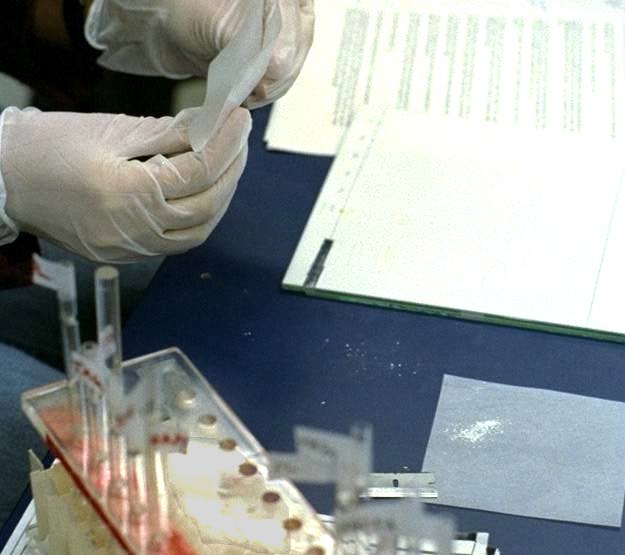
Handle
the plates carefully. Don?t leave any fingerprints. These will be shown after exposing to I2 vapour.
1. Using the template provided,
mark the plates with a sharp pencil.
2. Line the chamber with chromatography paper. Prepare 202 ml of solvent system (Hexane:Ether:Acetic
acid 60:40:1) in a 500 ml Erlenmeyer flask. Mix and pour ~150 ml into the chamber. Cover and let the chamber saturate while loading
the plates.
3. With a 10 ul capillary pipette, spot 1-2 ul of phospholipids standard onto the TLC plate, as shown. Make sure
the spot remains smaller than 4 mm in diameter. Move on to the other standards. After the spots have dried, repeat loading each standards
until you have loaded approx. 10 ul each. Also, load 10 ul of your lipid extract on one spot, and then the remainder of the extract
as a line (i.e., a series of spots).
4. Let dry the spots. Make sure that the loading area is above the solvent. Place the plates
in the chamber to develop.
5. Immediately close the cover and let run for approximately 30 min, until the solvent front has
reached the upper line.
6. Remove the plate and leave to dry in the rack in the fume hood. Discard the solvent in the waste
container provided, remove the chromatography paper and leave in the chamber. Leave the chamber in the fume hood to dry.
7.
Now place the plate in the iodine tank in the fume hood. You will see the lipids as yellow spots after about 5 min or so.
8.
Mark the edges of the spots with a pencil. Make a tracing on onion skin paper for a record.
9. Scrap off lipid fractions as shown,
place in weighing paper, fold and roll to grind the clumps.
10. Meanwhile prepare columns to elute the lipids from silica gel.
To do this insert a small amount of glass wool into a pasteur pipette, label the pipette and leave on the stand provided.
11.
Carefully transfer the silica gel having different lipids into appropriate columns. Keep 7 ml glass vials beneath the columns.
12.
Drip 1 ml of chloroform into each column except for the phospholipid and monoacylglycerol columns. To the latter, add >1 ml of
100 % methanol.
13. Shake the vials well. Place them in the fume hood evaporating set-up and evaporate off the solvent under a stream
of N2. (Ask the instructor before using the N2 tank).
14. Store the dry lipids under N2.
15. Label and leave the separated
lipid in the -20 C freezer until
transmethylation (day 2).
TLC Procedure, Educational Movie, Troubleshooting
and more.
Protocol 2. Brad's TLC protocol
Protocol 3. Immunostaining Thin Layer Chromatography of Glycolipids
Protocol 5. Lipid Analysis: Thin Layer Chromatography
Protocol 6. Thin Layer Chromatography
Web Guider
Ch 8.Immunohistoch / immunology
Ch 10.GC/MS, NMR and Proteomics